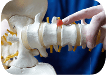
Lateral lumbar interbody fusion (XLIF) is a spinal fusion technique performed from the side of the body rather than from the back or through the abdomen. Spinal fusion procedures are performed for the relief of persistent pain in the lower back, the lumbar region of the spine. Interbody fusion refers to surgery in which an interverterbal disc is removed and the adjacent vertebrae are joined. The connection between the two vertebrae is accomplished through the use of a bone graft or through the insertion of bone morphogenetic protein, a manufactured substance also naturally found in the body.
XLIF can be used to treat nerve compression, disc degeneration, spondylothesis and other painful lower back conditions.
Benefits And Limitations Of XLIF
XLIF is different from other forms of fusion surgery because it uses a lateral, or side, approach to the spinal column. Approaches from the posterior, or back, necessitate the disturbance of muscles, nerves, blood vessels and ligaments in the back. Approaches from the anterior, or front, of the body pass through the abdominal muscles and come near the aorta and other vascular structures and urinary organs.
By approaching from the side of the body, the spine can be reached with minimal tissue damage and minimal risk. In addition, the side approach to the spine provides access to the damaged disc without requiring removal of any vertebral bone. Since the XLIF process is minimally invasive to the muscles and tissues at the site, complications are fewer and recovery is faster than with other fusion procedures.
XLIF, however, can only be performed on vertebrae that can be reached from the side and no more than two levels of the spine can generally be treated with this procedure. Also, XLIF is not performed to treat the lowest vertebrae on the spine. For these reasons, XLIF is not appropriate for all patients.
The XLIF Procedure
Once the incision is made, a probe is inserted to assist the surgeon in detecting compressed nerve roots and avoiding healthy nerve tissue. Dilators are used to separate and gently retract the muscles. An imaging device helps to ensure precision as the surgeon removes the injured disc, bone spurs and any other nearby debris. This restores room for the nerves that have been compressed.
A bone graft or bone morphogenetic protein is then inserted into the space that has been created. BMP has been approved by the FDA to stimulate bone growth in spinal fusion surgery. Once the surgeon has inserted the spacer that contains the graft material, being careful to avoid the spinal cord and adjacent nerves, metal plates, rods and screws are attached as needed. These instruments will hold the bones in place, maintaining spinal stability as the vertebrae grow together.
Once the XLIF procedure is complete, imaging is used again to confirm accurate placement. The surgeon then closes the incisions with sutures. Depending on the extent of spinal damage being repaired, the XLIF procedure takes between 1 and 3 hours.

